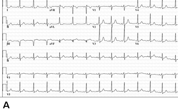
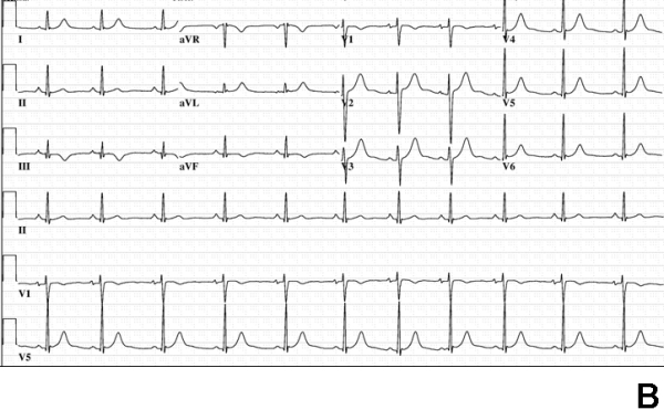
Figure 1.


Electrocardiograms at baseline (panel A) and postablation. Baseline ECG shows manifest preexcitation with suggestion of a right-sided pathway (positive delta wave in lead I, S wave larger than R wave in lead V1). Panel B shows absence of preexcitation following ablation in the left posterior region (see text for details).
