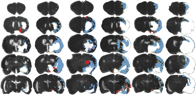
Figure 1.
Pattern of FMISO retention and infarction after 2-hour MCA occlusion with 24-hour survival. The areas of infarction only (red), infarction and [3H]FMISO retention (white), and [3H]FMISO retention without infarction (FMISO-defined penumbra, blue) are superimposed over images of the histologic sections. Coronal sections at five tissue planes are shown for experimental animals 1 to 6 in the corresponding columns, arranged by infarct volume. A concurrent sampling methodology was used to superimpose the areas of infarction and those of FMISO retention from autoradiographs of the same tissue slices. [3H]FMISO, hydrogen-3 fluoromisonidazole; MCA, middle cerebral artery.

