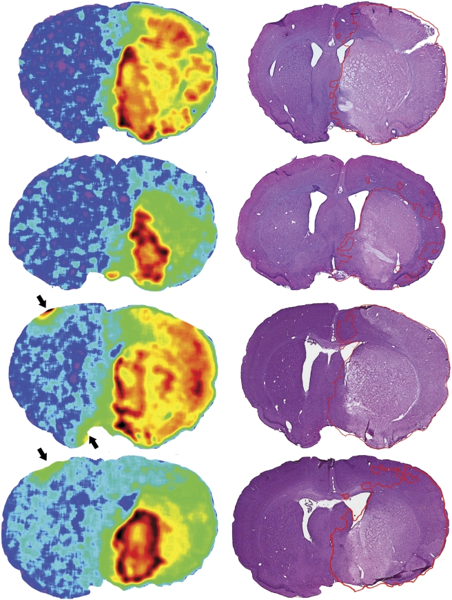
Figure 5.
FMISO retention in the infarct core. The autoradiograph and corresponding histology from a single tissue plane of each of the four animals with [3H]FMISO administered 6 hours after the onset of permanent MCA occlusion are shown. The histologic image has the outline of the FMISO retention superimposed, merely for illustrative purposes, because the threshold used was derived for 2-hour MCA occlusion and may overestimate the penumbra in permanent MCA occlusion (Spratt et al, 2009). Avid striatal retention is seen in all animals, corresponding to the regions of greatest H&E pallor. Arrows represent small areas of artefact caused by overlabelled fiducial marks, used to align histologic and radiologic images. [3H]FMISO, hydrogen-3 fluoromisonidazole; H&E, haematoxylin and eosin; MCA, middle cerebral artery.

