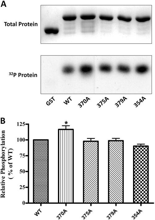
Fig. 2.
In vitro phosphorylation of GST-MOPr-CT by PKC. PKC-phosphorylated proteins (5 μg), including GST and GST-fused MOPr C terminus, were resolved by 12% SDS-PAGE. A, top, protein bands stained with Coomassie Blue; bottom, autoradiograph of the same gel. B, the phosphorylation of each protein was measured by scintillation counting and normalized to total protein. Results are expressed relative to phosphorylated wild type protein (100%). Data are presented as means ± S.E.M. (n = 6). *, P < 0.05 compared with WT (one-way ANOVA with Tukey post test).

