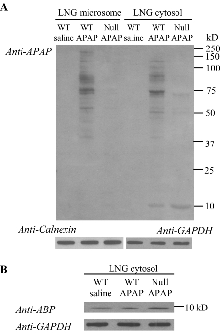
Fig. 7.
Immunoblot detection of APAP-protein adducts in the LNG of APAP-treated mice. Two-month-old male mice were fasted overnight before an injection of APAP (at 400 mg/kg i.p.); mice in the control group were treated with saline. Microsomal and cytosol preparations were obtained from the pooled LNGs of four mice; tissues were obtained at 2 h after the APAP treatment. Microsomal and cytosol (A) samples (20 and 30 μg of protein, respectively, in each lane), from either B6 WT or Cyp2a5-null (Null) mice, were analyzed on 10% SDS-polyacrylamide gels, and APAP adducts were detected on immunoblots by using a polyclonal anti-APAP antiserum. B, the levels of ABP protein were determined on a separate blot for the same cytosol samples that were used in A to demonstrate induction of the ABP protein in the Cyp2a5-null mice. The levels of calnexin (for microsomes, A) and GAPDH (for cytosol, A and B) were also determined, after stripping of the blot to remove the anti-APAP or anti-ABP antibodies, for demonstration of equal protein loading among the three samples. Densitometric analysis (not shown) indicated a ∼2.5-fold higher level of APAP-ABP (A) and a 2.8-fold higher level of ABP (B) in the LNG cytosol from APAP-treated Cyp2a5-null mice, compared with APAP-treated WT mice.

