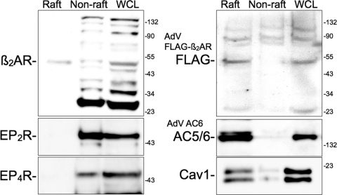
Fig. 6.
Immunoblot analysis for GPCRs and AC6 expressed in lipid raft and nonraft fractions from hBSMC. Cells were fractionated using a nondetergent method and separated by sucrose density centrifugation (see Materials and Methods). Buoyant fractions (Raft) and nonbuoyant “heavy” fractions (Non-raft) were pooled and loaded along with whole-cell lysates (WCL) into individual lanes and separated by SDS-PAGE. Blots are labeled with the primary antibody at the approximate molecular weight of the expected immunoreactive band. In some studies, hBSMC were incubated for 24 h with recombinant adenoviruses expressing either FLAG-β2AR or AC6. Images shown are representative of three to four experiments. Cav1, caveolin-1.

