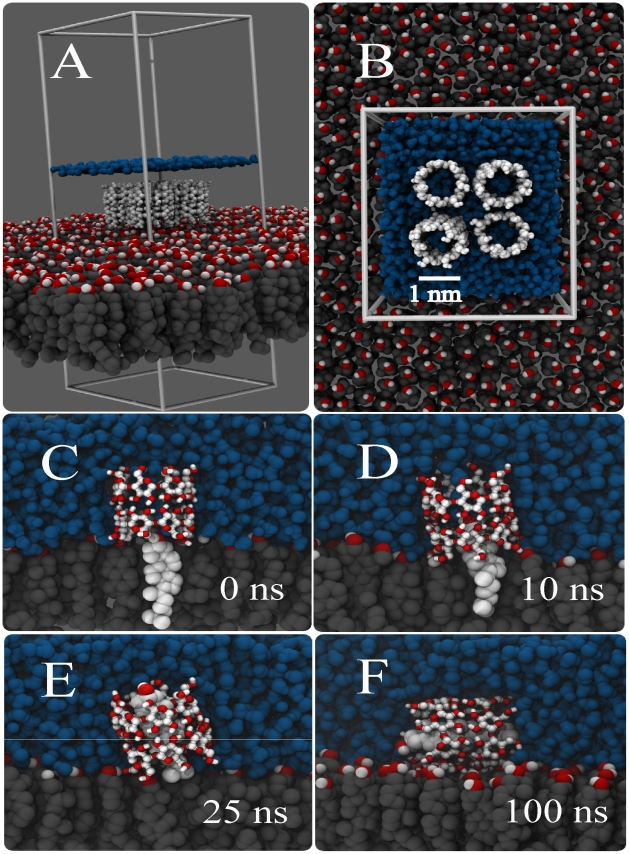
Figure 1. Spontaneous extraction of cholesterol by a cyclodextrin dimer.
Panels (A,B) show the initial system set-up with four CD dimers sitting on top of a pure cholesterol monolayer, panels C–F show the time evolution of the extraction of cholesterol by one of the CD dimers. At 0 ns the cholesterol is still inside the monolayer (C), at 10 ns the upper part is sucked in (D), and after 25 ns the cholesterol is almost fully inside apart from the tail (E). Tilting of the complex after 100 ns completes the extraction process (F). Color code; cholesterol body: grey, cholesterol head: red-white, CDs: white and red, water: blue (In panel A the level of the water layer is only depicted by a blue line).

