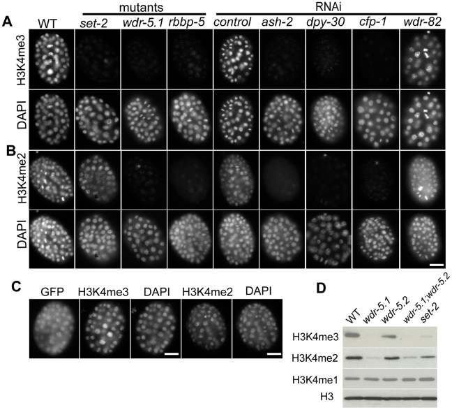
Figure 1. Set1/MLL complex components are largely responsible for H3K4me2/3 in embryos.
Embryos were dissected from WT, mutant, or RNAi treated adult eri-1(mg366; enhanced RNAi) animals. RNAi mediated knockdown was used for ash-2, dpy-30, cfp-1 and wdr-82. Embryos were probed with rabbit anti-H3K4me3 (Panels in A) or mouse anti-H3K4me2 monoclonal antibodies (Panels in B); DNA was counter-stained with DAPI. Exposure times were the same for each condition. Scale bars represent 10um. (A and B) H3K4me3 and H3K4me2 are present in chromatin of wild-type embryos, but both are dramatically decreased in chromatin of embryos from wdr-5.1 and rbbp-5 mutants, and embryos from ash2, dpy-30, or cfp-1 RNAi treated mothers. H3K4me3, but not H3K4me2, is reduced in set-2(tm1630) mutant embryos. RNAi mediated knockdown of wdr-82 did not affect either H3K4me3 or H3K4me2. (C) A wdr-5.1::GFP transgene rescues H3K4 methylation in the wdr-5.1(ok1417) mutant. wdr-5.1(ok1417);wdr-5.1::GFP transgenic embryos were probed with antibodies against GFP and H3K4me3/2 as indicated. H3K4me3/2 were restored in the chromatin of early embryos in which WDR-5.1:GFP expression was detected. (D) Western blot of lysates from mixed stage wdr-5.1(ok1417), wdr-5.2(ok1444), wdr-5.1;wdr-5.2 double mutant, and set-1(tm1630) mutant embryos. The blots were probed with anti-histone H3, anti-H3K4me1, mouse monoclonal anti-H3K4me2 or rabbit polyclonal anti-H3K4me3 antibodies, as indicated. H3K4me3/2 levels were dramatically decreased in strains with the wdr-5.1(ok1417) allele, whereas H3K4me3 levels were more strongly depleted than H3K4me2 in set-2(tm1630). H3K4me1 levels were not noticeably affected in any mutant strain tested.

