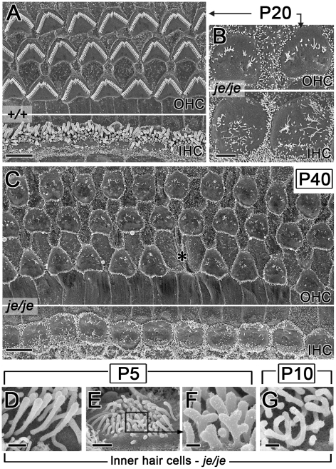
Figure 3. Advanced stereociliary degeneration on cochlear hair cells and peculiar stereociliary defects on inner hair cells.
(A–C) Stereociliary degeneration involving shortening and disappearance (see Figure 2) progresses for the outer hair cells (OHC) and the inner hair cells (IHC) of je/je mice. By P20 and P40, only sparse, disorganized collections of small stereociliary remnants remain (B,C). The middle region of the cochlea is shown. Panel A shows the corresponding region in P20 +/+ mice. Asterisk in C, position missing an outer hair cell. (D–G) Higher magnification reveals peculiar defects in je/je mice at P5 and P10, including bulbous tips for inner hair cell stereocilia in the tallest row (D) and branched, antler-like surface projections on the opposite (neural) face of the collection (E–G). F is a higher-magnification view of the box in E. Scale bars, 5 µm (A,C), 2 µm (B), 0.5 µm (D), 1 µm (E) or 0.2 µm (F,G).

