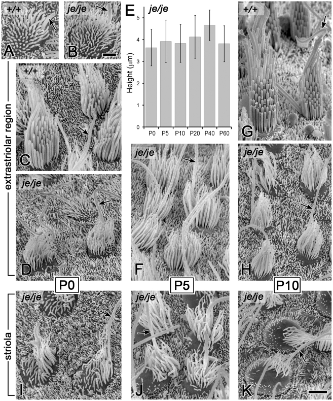
Figure 5. Morphogenesis defects and degeneration of vestibular hair cell stereocilia in utricular maculae of je/je mice.
Extrastriolar and striolar regions of utricular maculae are shown (kinocilium, arrow). (A,B) At P0 the surface projections of immature utricular hair cells in je/je mice (B) look similar to those in +/+ mice (A) in length and length-gradation, but those in je/je mice appear thinner. (C,G) During stereociliary morphogenesis in +/+ mice, stereocilia increase in diameter and undergo differential elongation to form a relatively tall staircase. (D–F,H–K) Stereociliary morphogenesis is significantly impaired in je/je mice. Stereocilia are much thinner and appear to taper gradually in the proximal-to-distal direction, and there is little or no differential elongation to form a staircase. Additionally, in the striolar region, progressive stereociliary degeneration is evident: stereocilia become crooked and appear to collapse by buckling in their proximal segment (J,K). In contrast, stereocilia in the extrastriolar region attain a length that is approximately one-half that of the tallest stereocilia observed on the extrastriolar hair cells of +/+ mice and remain upright for a longer period of time (D,F,H). The average height of the stereociliary collection in je/je mice changes little between P0 and P60 (E). Examples of stereocilia on the striolar hair cells of +/+ mice are shown in Figure S3A and S3C. Scale bars, 1 µm (A,B) or 2 µm (C,D,F–K).

