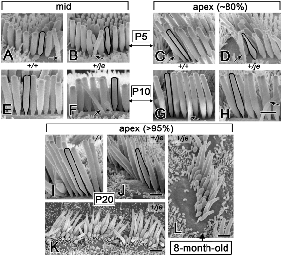
Figure 13. Tapering of stereocilia on the inner hair cells of +/je mice.
(A–H) Stereocilia on inner hair cells in the middle and apical cochlear regions of +/je mice show obvious tapering along their proximal-distal axis (B,D). This tapered segment becomes mostly (F) or partly (H) filled in by P10. The tapering is not observed for the stereocilia of +/+ mice (A,E,C,G). Black outlines highlight examples (kinocilium, arrow). (I-L) In the extreme apex (>95% from base), proximal-distal tapering of stereocilia is evident in +/je mice at P20 (J,K), but not in +/+ control mice (I), and can still be seen at 8 months (L). Scale bars, 1 µm (A-J,L) or 2 µm (K).

