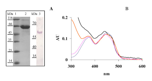
Figure 2.
Spectral analysis of purified B. breve MCRA protein. SDS PAGE analysis of B. breve MCRA protein purified on Ni-NTA column. Lane 1, molecular weight marker, lane 2, B. breve MCRA protein of apparent MW of 66 kDa, lane 3, immunoblot with anti-His tag antibody shows the absence of significant proteolytic degradation in purified B. breve MCRA. B UV-Vis spectrum of B. breve MCRA protein (black) and the supernatant after heat precipitation (red), showing flavin-like absorbance. For the reference spectra of 10 μM FAD (blue) and FMN (magenta) are shown.

