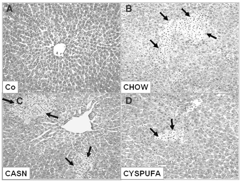
Figure 3.

Representative hepatic sections stained with hematoxylin and eosin 18 hours after (A) saline and (B–D) lipopolysaccharide (LPS) administration. (A) Slide from representative rat injected with saline (Co) shows normal hepatic structure. (B–D) Hepatic section from representative rat that consumed CHOW (B) 18 hours after LPS injection shows severe multifocal necrosis, infiltration of inflammatory cells, and necrosis (arrows). (C) CASN diet and (D) CYSPUFA diet show mild focal necrosis with less severe lesions.
