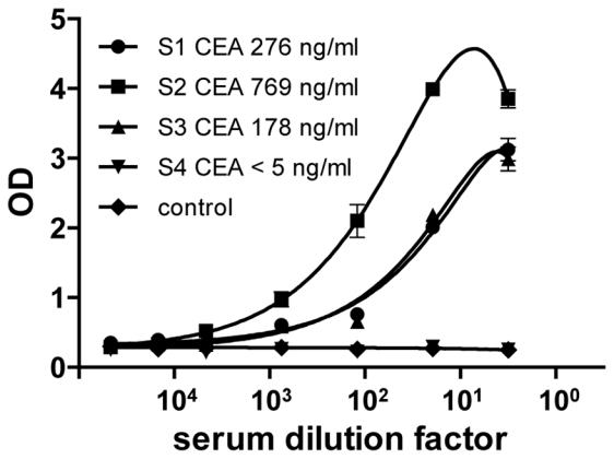
Figure 5. Immobilization of in vivo biotinylated sdAbs allows a sensitive detection of CEA in patient sera using a bead assay.

Streptavidin beads were coated with bacterial lysates (0.5 μl/wells) containing in vivo biotinylated (●,■, ▼, ▲) or unbiotinylated (◆) anti-CEA sdAbs and incubated with serial dilutions of patient sera (●: S1 CEA 276 ng/ml, ■: S2 CEA 769 ng/ml, ▲: S3 CEA 178 ng/ml, ▼: S4 CEA < 5ng/ml). The captured antigen was detected with a mouse anti-CEA antibody (35A7) followed by a goat anti-mouse HRP-conjugated mAb. Standard deviation represents two experiments performed in triplicates.
