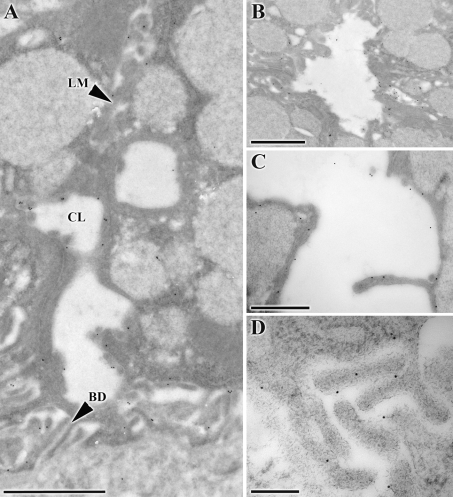
Fig. 4.
Gold labeling of AQP5 in the SMG of adult animals (P60). A Overview of AQP5 gold staining in the basal membrane, membranes of intercellular canaliculi, as well as the lateral membrane (LM). CL canalicular lumen, BD basal digits (scale bar, 1 μm). B Gold labeling of AQP5 was detected in the apical membrane of acinar cells (scale bar, 1 μm). C Longitudinal section of an intercellular canaliculus showing gold labeling of AQP5 in the membrane (scale bar, 0.5 μm). D AQP5 was localized in the basal membrane in areas where digits could be seen, while no AQP5 was detectable in the basement membrane (scale bar 0.2 μm). Non-linear adjustments were applied to entire images

