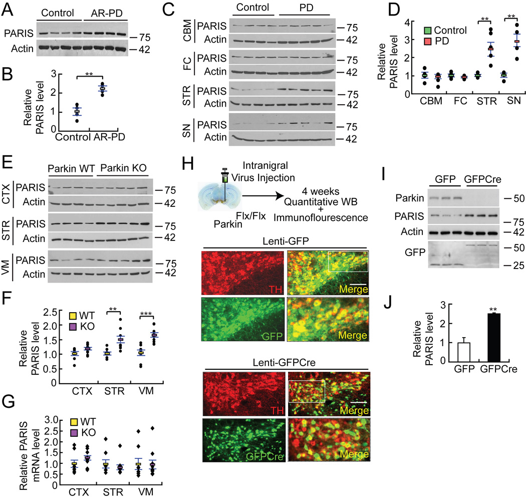
Figure 4. PARIS accumulates in AR-PD, sporadic PD and in animal models of parkin inactivation.
(A) Immunoblot analysis of PARIS and β-actin in cingulate cortex from age-matched controls and AR-PD patient brains with parkin mutations
(B) Quantitation of the immunoblots in panel A normalized to β-actin, n = 4.
(C) PARIS levels in cerebellum (CBM), frontal cortex (FC), striatum (STR) and SN of sporadic PD patient brains compared to age-matched controls.
(D) Relative PARIS levels normalized to β-actin in panel B, Controls n = 4; PD n = 5.
(E) Immunoblot analysis of PARIS in cortex (CTX), STR and ventral midbrain (VM) from WT and parkin exon 7 KO 18–24 month old mice.
(F) Relative protein levels of PARIS normalized to β-actin for panel E, WT n = 9; parkin KO n = 10.
(G) PARIS mRNA levels in indicated brain regions from WT and parkin exon 7 KO 18–24 month old mice.
(H) Top panel, experimental illustration of stereotaxic intranigral virus injection. Bottom panels, immunofluorescent images of TH (red), GFP (green) and merged (yellow) in exon 7 floxed parkin mice (parkinFlx/Flx) after stereotactic delivery of Lenti-GFP or Lenti-GFPCre into the SNpc. 84.9±1.9% and 78.1±2.6% of TH neurons express GFP and GFPCre, respectively, n = 3 per group. Enlarged images in the right bottom panels were taken from the white rectangle region from the merged images of Lenti-GFPCre and Lenti-GFP, bar = 100 µm.
(I) Immunoblot analysis of parkin, PARIS, actin and GFP 4 weeks after intranigral Lenti-GFPCre or Lenti-GFP injection into parkinFlx/Flx mice.
(J) Relative protein levels of PARIS normalized to β-actin for panel I. Data = mean ± S.E.M., *p < 0.05, **p < 0.01 and ***p < 0.001, unpaired two-tailed Student’s t-test (B, J) and ANOVA test with Student-Newman-Keuls post-hoc analysis (D, F, G).
See also Table S1 and S2.

