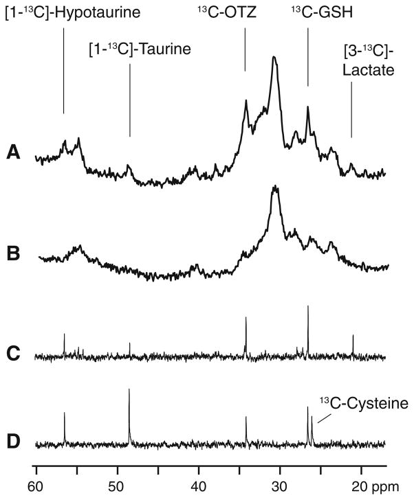
Fig. 3.
a A portion of the in vivo 13C magnetic resonance spectrum showing signals originating from subcutaneous fat and brain from a rat after 20 h of 13C-OTZ infusion at a total dose of 1,300 mg/kg. b The natural abundance in vivo 13C magnetic resonance spectrum showing signal originating from subcutaneous fat and brain from a control rat. c A portion of the high-resolution 13C magnetic resonance spectrum of the acid extract of brain tissue from the same rat used in a. d A portion of the high-resolution 13C magnetic resonance spectrum of the acid extract of liver tissue from the same rat used in a and spiked with a standard solution of [3-13C]-cysteine. In addition to resonances identified in Fig. 2, [3-13C]-lactate is observed at 21.0 ppm, [1-13C]-taurine at 48.5 ppm, [1-13C]-hypotaurine at 56.5 ppm. The liver sample shows an additional resonance from the added [3-13C]-cysteine at 26.1 ppm

