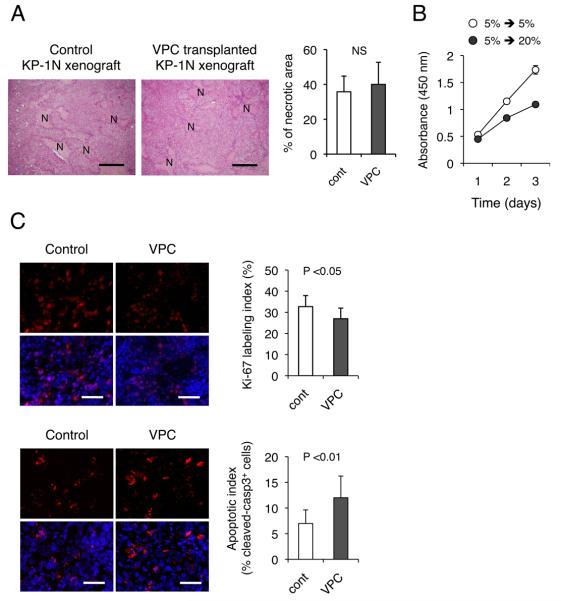
Figure 4. Transplantation of ex vivo cultured VPC impairs growth of KP-1N xenografts.
(A) KP-1N xenografts were treated with or without intravenous administration of cultured VPCs (5 ×10⁵ cells; twice) and mice were sacrificed 2 weeks after the procedure. H-E stainings for xenograft sections are shown and % necrotic area (N) was measured. Scale bars; 200 μm. (B) KP-1N cells adapted to chronic hypoxia (5 % O2 in hypoxia workstation INVIVO400) were culture wither in normoxic (20 % O2) or hypoxic conditions (5 % O2) for 3 days. Cell growth was quantified by WST-8 assay. (C) Xenograft tissues were stained with anti-Ki-67 (upper panel) and anti-cleaved caspase-3 (lower panel). Quantification of proliferating/apoptotic is shown as percent positive cells in 5 viable fields from 10 sections. Scale bars; 100 μm. Proliferation/Apoptotic index are shown (mean ± SEM).

