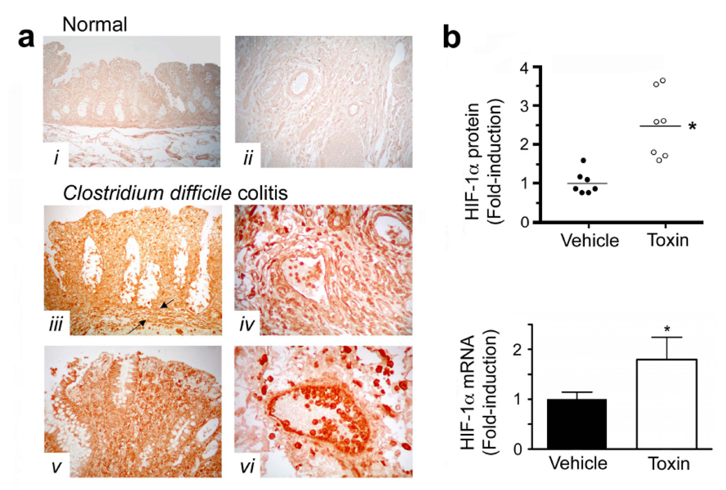
Figure 1.
C. difficile colitis is associated with increased HIF-1α expression. (a) Tissue sections were stained for HIF-1α protein using a mouse anti-HIF-1α monoclonal antibody (NB100-105). Immunolocalization of HIF-1α was determined in human colonic tissue obtained from patients (surgical resections): (i– ii), HIF-1α staining was absent from normal human colonic tissue obtained from biopsy during colon cancer screening or normal resection margins for colon cancer; (iii–vi), intense HIF-1α staining was noted in colonic tissues with C. difficile-associated pathology. The mucosa, submucosa and muscularis with discrete staining identified in inflammatory cells, epithelial cells, endothelial cells, as well as smooth muscle cells of the muscularis mucosa (between arrows), the intimal vasculature and the muscularis. Standard immunohistochemical controls (e.g. omission of mouse anti-HIF-1α antibody) were negative. (b) HIF-1α protein and mRNA are induced in non-inflamed, human colonic biopsies following exposure to C.difficile toxin (50µg/mL; 3h). Two biopsies (taken side by side) were collected from healthy individuals and immediately incubated with either toxin or vehicle solution (matched volume of sterile BHI media). All values are mean ± S.E.M., (n=14). * denotes p<0.05.

