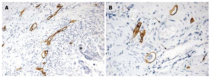
Figure 2.

Morphological features of D2-40 positive lymphatic vessels in colorectal cancer. A: Positive D2-40 stained lymphatic vessels with thin walls and irregular shapes in peritumoral area (asterisk, × 100); B: Positive D2-40 stained lymphatic vessel containing tumor emboli within tumor mass (arrow head) and D2-40-negative erythrocytes (black arrows, × 200).
