
Fig. 2.
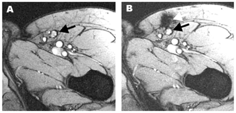
Monitoring of the accuracy of delivery of SPIO-labeled DCs using MRI. Black arrow denotes the inguinal lymph node (A) and white arrow indicates that the location of labeled DCs not in the lymph node but in the perinodular fat (B). Reproduced from ref. 81 with permission.