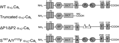
Fig. 1.
Schematic depicting the domain structure of wild-type (WT) L-type voltage-gated Cav1.2 calcium channel (CaL) α1C-subunit and mutant α1C-CaL constructs used in this study. I–IV, the four transmembrane repeat of α1C. EF and DCT, EF hand motif and the distal COOH terminus inhibitory domain; P, proline-rich domain; truncated α1C-CaL, WT α1C-CaL missing the distal COOH-terminal 280 amino acids; ΔP1/ΔP2 α1C-CaL, WT α1C-CaL missing the two proline-rich domains (aa 1955–1959; aa 1967–1973); S1901A/Y2122F α1C-CaL, two single-point mutations at serine (S1901A) and tyrosine (Y2122F) in full-length neuronal WT-CaL.

