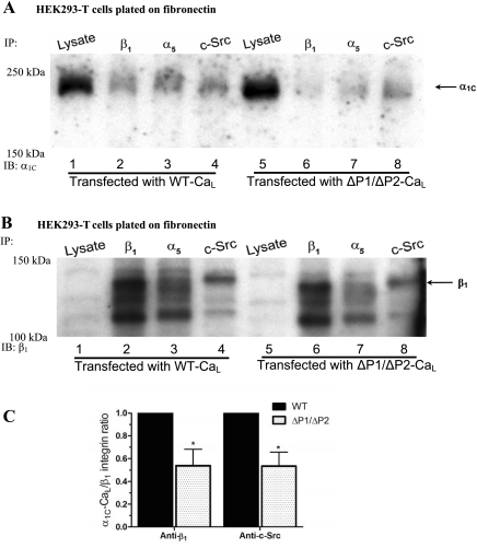
Fig. 6.
Requirement of proline-rich domains (PRDs) in the α1C-CaL COOH terminus for α1C-CaL association with β1-integrin or c-Src. A: representative blot comparing WT α1C-CaL and ΔP1/ΔP2 α1C-CaL immunoprecipitated with anti-c-Src, anti-α5-integrin, or anti-β1-integrin Ab and probed for α1C. Relatively lower amounts of α1C-CaL appear to be pulled down by anti-c-Src or anti-β1-integrin Ab (lanes 8 and 6, respectively) in cells expressing ΔP1/ΔP2 α1C-CaL, compared with WT α1C-CaL. B: the same blot in A after stripping and reprobing for β1-integrin. C: summary graph showing the relative levels of immunoprecipitated α1C in WT- or ΔP1/ΔP2-CaL. Values were obtained as described in methods and are based on the average of at least three experiments. *P < 0.05 vs. WT + anti-c-Src Ab or WT + anti-β1-integrin Ab.

