Fig. 7.
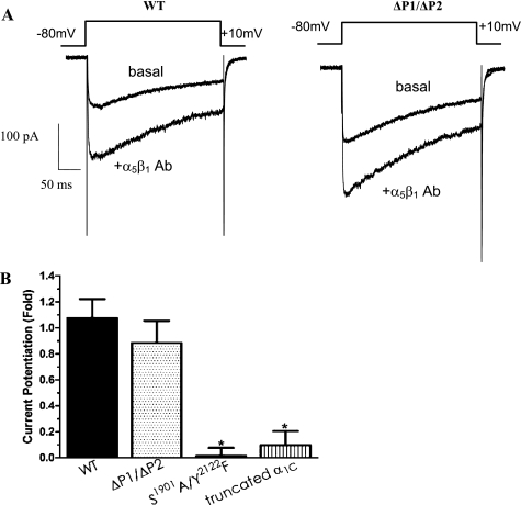
Electrophysiological protocols to determine the effects of COOH terminus-specific regions on modulation of CaL current by α5β1-integrin. A: representative recordings of whole cell CaL current from HEK293-T cells expressing WT-CaL (left) or ΔP1/ΔP2-CaL (right). Top traces in both panels show the amount of basal current in cells expressing WT- or ΔP1/ΔP2-CaL, and bottom traces show the current potentiation by α5β1-integrin Ab (10 μg/ml). No substantial difference in the relative amount of CaL current potentiation was observed in cells expressing ΔP1/ΔP2-CaL, compared with WT-CaL. B: summary graph showing peak current potentiation by α5β1-integrin in WT-CaL, truncated α1C-CaL, or ΔP1/ΔP2-CaL. CaL current potentiated by α5β1-integrin Ab was significantly attenuated in truncated α1C-CaL or in S1901A/Y2122F-CaL, but not in ΔP1/ΔP2-CaL. Number of cells recorded is as follows: WT-CaL, n = 18; n = 8 in cells expressing ΔP1/ΔP2-CaL, S1901A/Y2122F-CaL, or truncated α1C-CaL. *P < 0.05 vs. WT-CaL.

