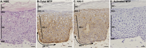
Fig. 1.
Lack of matriptase activation in normal epidermis. Samples of normal human skin were used to prepare frozen sections that were stained with hematoxylin and eosin (H & E; A) or immunostained with monoclonal antibodies to detect total matriptase M32 (total MTP; B), total hepatocyte growth factor activator inhibitor (HAI)-1 M19 (HAI-1; C), or activated matriptase M69 (activated MTP; D). Mouse IgG was used as the negative control (not shown). SB, stratum basale; SS, stratum spinosum; SG, stratum granulosum; SC, stratum corneum. Staining intensity is expressed as ++ for strong positive, + for positive, +/− for weak positive, and − for negative. Bars = 25 μm.

