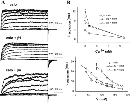
Fig. 4.
Activation of Slo, dependent on voltage and Ca2+, is slowed by β4 and β1. A: current traces of inside-out macropatches from oocytes injected with cSlo, cSlo and β1, and cSlo and β4 cRNA subjected to a step voltage protocol. Patches were held at −100 mV and stepped to a range of voltages varying from −80 to +180 mV before stepping back to −100 mV (1 μM intracellular Ca2+). β4 slows activation more than β1 (cSlo, n = 15; cSlo + β1, n = 12; cSlo + β4, n = 20). B: activation time constants (τactivation) as a function of Ca2+ (with voltage stepped from −100 to +60 mV, top) and voltage (with intracellular Ca2+ held at 1 μM, bottom). As is evident, higher voltage and Ca2+ both result in shortened times of activation.

