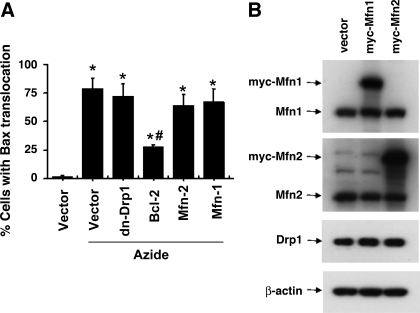
Fig. 2.
DN-Drp1, Mfn1, and Mfn2 do not block Bax translocation to mitochondria during azide treatment. A: Bax translocation analyzed by immunofluorescence staining. HeLa cells were cotransfected with MitoRed and one indicated plasmid (empty vector, dn-Drp-1, Bcl-2, Mfn1, or Mfn2). After azide treatment, the cells were fixed for Bax immunofluorescence staining (labeled by green FITC) and examined to count the cells with Bax translocation to mitochondria. Data are expressed as means ± SD (n = 4); *P < 0.01 vs. control; #P < 0.01 vs. azide treated vector-transfected group. B: Myc-Mfn1 and Myc-Mfn2 expression after transfection. Whole cell lysate was collected for immunoblot analysis using specific antibodies to Mfn1, Mfn2, Drp1, and β-actin.

