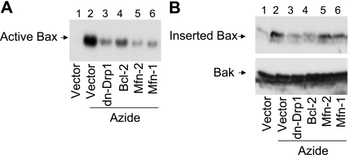
Fig. 3.
DN-Drp1, Mfn1, and Mfn2 suppress Bax activation and insertion in mitochondria membrane. HeLa cells were transfected with Mfn1, Mfn2, dn-Drp1, Bcl-2, or control empty vector. The cells were then subjected to 3 h of 10 mM azide treatment in glucose-free buffer. A: active Bax. Cells lysate was collected for immunoprecipitation with an antibody that was specific to active Bax. The precipitate was finally analyzed by immunoblotting of Bax. B: Bax insertion. Membrane fraction containing mitochondria was collected from the cells and incubated for 30 min with an alkaline (pH 11.5) solution. The mitochondrial fraction was then collected by centrifugation for analysis of remaining Bax by immunoblot analysis. Bak was also analyzed to verify that inserted proteins were not stripped off from mitochondrial membrane by the alkali incubation.

