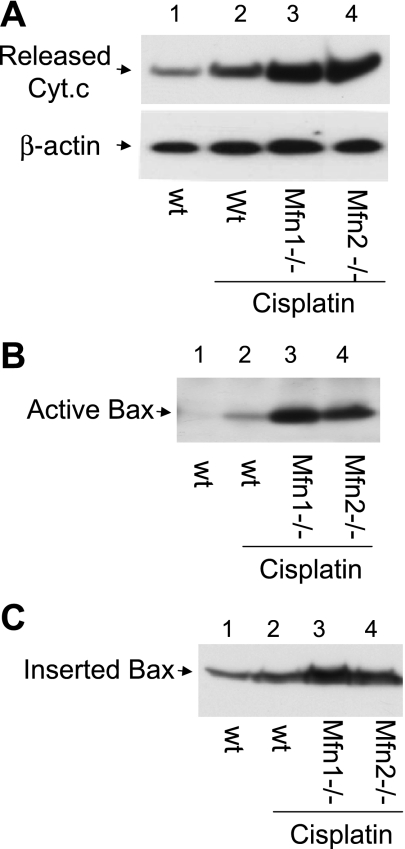
Fig. 7.
Mfn1 or Mfn2-null cells are more sensitive to Cyt c release, Bax activation, and insertion. wt, Mfn1-null, and Mfn2-null MEFs were incubated with 20 μM cisplatin for 24 h. A: Cyt c release. Cells were permeabilized with low concentration digitonin to collect cytosolic fraction for immunoblot analysis to detect Cyt c that had been released into cytosol during cisplatin treatment. B: Bax activation. Cells were lysed with the CHAPS buffer. The lysate was subjected to immunoprecipitation using the antibody specific for active Bax. The resultant immunoprecipitates were analyzed for Bax by immunoblot analysis. C: Bax insertion. Cells were permeabilized with digitonin to release cytosol and collect the membrane fraction with mitochondria, which was subjected to alkaline incubation as described in materials and methods. After alkaline treatment, Bax remaining in the membrane fraction was analyzed by immunoblot analysis.

