Fig. 3.
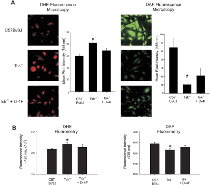
Effects of D-4F on·NO and O2·− balance in endothelial cell cultured on microfibrils. A, left: representative superoxide anion (O2·)-dependent hydroethidine (DHE) fluorescence images of endothelial cells (EC) cultured on different microfibril preparations. The levels of O2·− are low in EC cultured on microfibrils isolated from C57BL/6J mice (top). In contrast, O2·− levels in EC cultured on microfibrils isolated from Tsk−/+ mice are increased (center). EC cultured on microfibrils isolated from D-4F-treated Tsk−/+ mice (bottom) generate less O2·− than EC on Tsk−/+ microfibrils (center) but not as low as EC cultured on microfibrils from C57BL/6J mice (top). A, right: representative amino-5-methylamino-2′7′diaminofluorescein (DAF)-nitric oxide (·NO) fluorescence images of EC cultured on different microfibril preparations. EC cultures maintained on microfibrils isolated from C57BL/6J mice generate high levels of intracellular·NO based on intense green fluorescence of DAF-NO (top). EC cultures maintained on microfibrils isolated from Tsk−/+ mice generate low levels of intracellular·NO based on faint DAF-NO fluorescence (center). EC cultured on microfibrils isolated from D-4F-treated Tsk−/+ mice generate levels of DAF-NO that are higher (bottom) than EC cultured on Tsk−/+ microfibrils (center) but not as high as when EC are cultured on microfibrils from C57BL/6J mice (top). Bar graphs depict fluorescence intensity of 60 images for each group and treatment. Tsk−/+ is significantly different (#P < 0.05) from all the other groups. Tsk−/+ is significantly different (*P < 0.05) from C57BL/6J group. B: fluorescence intensity measured with microplate reader shows similar results as in A.

