Fig. 1.
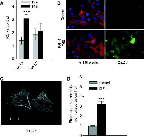
Insulin-like growth factor-I (IGF-I) upregulates T-type channels in cultured rat pulmonary artery smooth muscle cells (PASMCs). A: rat PASMCs were serum starved for 2–3 days before IGF-I stimulation. After 24 and 48 h in IGF-I, PASMCs were harvested for RNA purification and quantitative RT-PCR. RQ represents the relative quantification vs. time-matched control, unstimulated cells and is calculated based on the ΔΔCt (comparative threshold cycle) method, using 18S rRNA as endogenous control. Values are means ± SE. Statistical significance is calculated using paired t-test on dCt values. ***P < 0.001. B: immunostain of α-smooth muscle (α-SM) actin (SM-22, red), voltage-gated calcium channel (Cav) 3.1 (green), and nuclei counterstained with 4,6-diamidino-2-phenylindole (DAPI; blue) in unstimulated (control) and 48-h IGF-I stimulated PASMCs. Scale bar = 50 μM. C: confocal image of Cav3.1 immunostain in 48-h IGF-I-treated PASMCs (as in B); scale bar represents 20 μm. D: averaged fluorescence intensity of Cav3.1-immunostained PASMCs with 48-h IGF-I treatment, normalized to control untreated PASMCs (as in B). Values are means ± SE. ***P < 0.001 for IGF-I (n = 61) vs. control (n = 41).

