Figure 1.
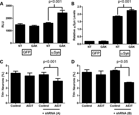
Potentiation of α-synuclein-mediated toxicity in cell culture systems with reduced GAK expression. (A and B) HEK 293 cells were transiently transfected with CMV-GFP, CMV-α-synuclein and either non-targeted (NT) or GAK-targeted pools of siRNA. (A) Toxicity was assessed by release of adenylate kinase into the media 72 h after transfection. Enzymatic release was detected by luciferase reaction and quantified by relative luminescent units (RLU) compared with controls (Toxilight). (B) α-Synuclein protein levels in siRNA-treated cell lysates were quantified by ELISA. (C and D) Primary midbrain cultures were transduced with A53T adenovirus (MOI = 3) with or without lentivirus encoding either (C) GAK shRNA ‘A’ (MOI = 3) or (D) GAK shRNA ‘B’ (MOI = 1). Control cells were incubated in the absence of virus. Dopaminergic cell viability was determined by staining with antibodies specific for MAP2 and TH and is expressed as the percentage of MAP2-positive neurons that were also TH-positive.

