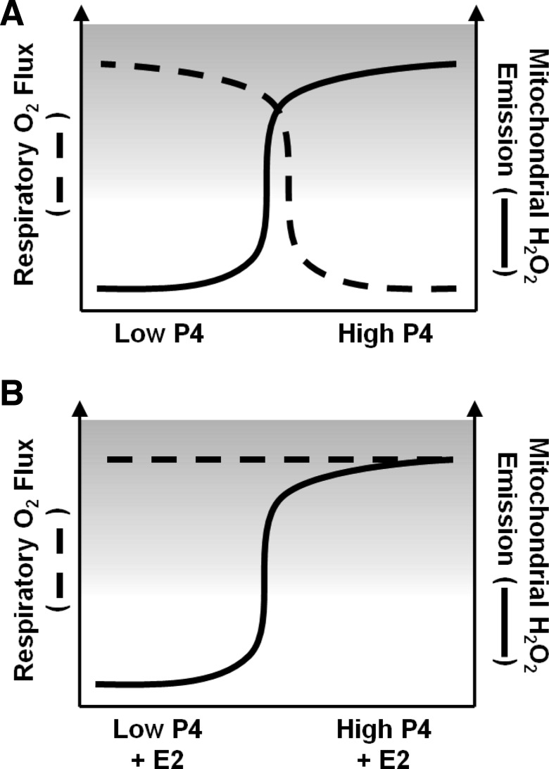
Fig. 5.
Diagrammatic summation of results and proposed effects of E2 and P4 on skeletal muscle mitochondrial function and insulin sensitivity. Both E2 and P4 are elevated during the luteal phase relative to the early follicular phase of the female menstrual cycle. Results of the current study support a model whereby elevated P4, whether alone (A) or in the presence of high E2 (B), increases the propensity for mEH2O2 in skeletal muscle of women. P4-associated inhibition of skeletal muscle Jo2 flux (A) is not observed when high E2 is present (B). Given that a reduction in insulin sensitivity has been reported for women in the luteal phase, and considered in the context of the observed increase in mEH2O2 with insulin resistance, results of the current study also suggest that the rise in P4 that accompanies the luteal phase may lead to insulin resistance via an increase in mEH2O2.

