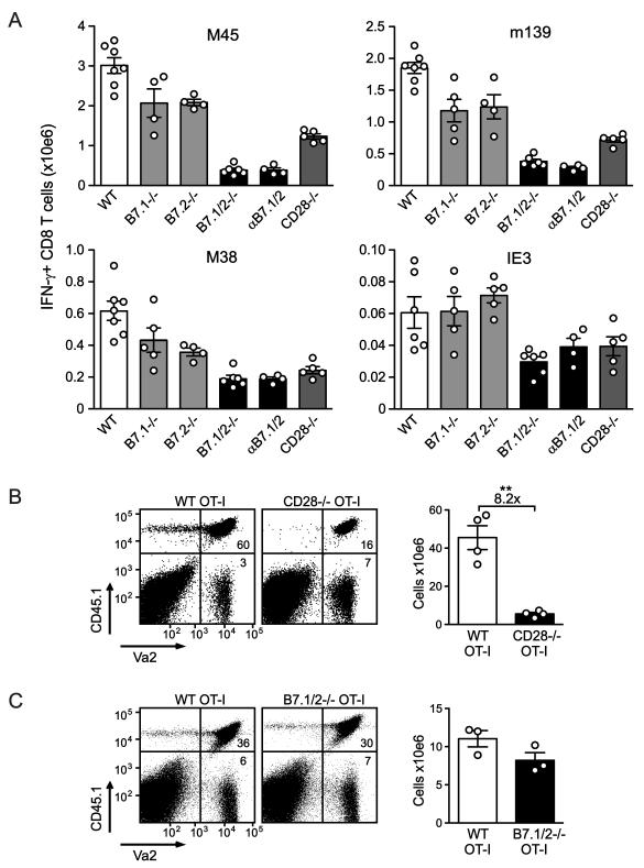
Figure 2. Both B7.1 and B7.2 promote initial expansion of MCMV-specific CD8 T cells.
(A) WT, B7.1−/−, B7.2−/−, B7.1/2−/−, CD28−/− and WT mice treated with blocking B7.1 and B7.2 antibodies (αB7.1/2) were infected with MCMV and 8 days later the virus-specific CD8 T cell response was analyzed by intracellular cytokine staining after restimulation with the indicated class I epitopes. Graphs show the total number of IFN-γ+ CD8 T cells in the spleen. (B) 1 × 10⁴ congenic (CD45.1) WT OVA-specific TCR transgenic CD8 T cells (OT-I) or CD28−/− OT-I cells or (C) 1 × 10³ CD45.1 WT OT-I or B7.1/2−/− OT-I cells were adoptively transferred into WT (CD45.2) mice that were subsequently infected with MCMV-OVA. The number of transgenic T cells was determined in the spleen 8 days following transfer. Statistical significance was determined by Student’s t test (** indicates p<0.005). Fold difference is indicated. All bar graphs in this figure are shown as means with standard errors and each symbol (○) represents an individual mouse. Data shown are representative of two independent experiments.

