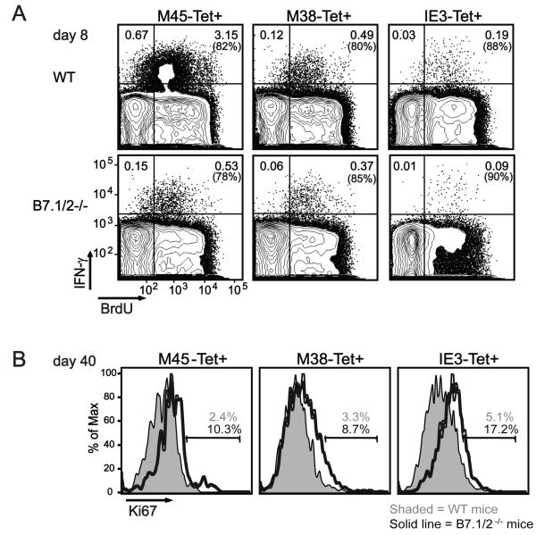
Figure 6. Increased proliferation of MCMV-specific CD8 T cells in mice lacking B7-CD28 signaling during persistent MCMV infection.
WT and B7.1/2−/− mice were infected with MCMV. (A) At day 8 post-infection BrdU staining was performed after in vivo labeling with BrdU (1 mg/ml in drinking water) for 7 days in combination with intracellular cytokine staining after restimulating splenocytes with the indicated class I epitopes. Plots are gated on CD8 T cells and show BrdU expression versus IFN-γ reactivity. (B) At day 40 post-infection the intracellular expression of the cell cycle marker Ki67 was analyzed on M45-, M38- or IE3-tetramer positive (Tet+) CD8 T cells by flow cytometry. Histograms are gated on splenic CD8 Tet+ cells. Data shown are representative of two independent experiments.

