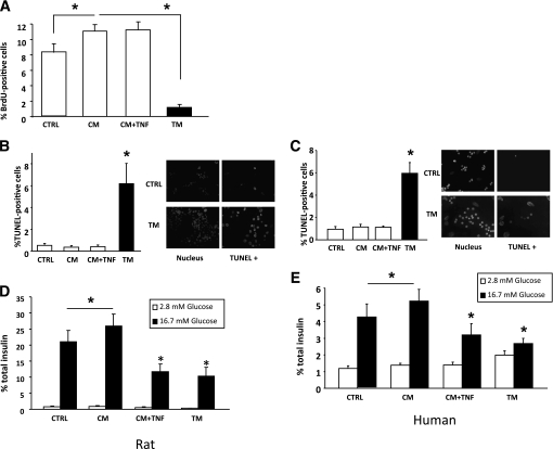
FIG. 4.
Effect of conditioned medium on primary β-cell proliferation, survival, and GSIS. Conditioned medium was obtained by culturing human myotubes for 24 h with (TM) or without (CM) 20 ng/mL TNF-α. Additional control conditions were as follows: CTRL, culture medium not previously exposed to skeletal muscle cells; CM + TNF, 20 ng/mL TNF-α added to CM immediately before exposure to β-cells. A: Proliferation of rat primary β-cells measured by BrdU incorporation. Cells were grown under standard culture conditions (20% FCS, 11.2 mmol/L glucose) and treated for 48 h with the different conditioned media; BrdU was added for the last 24 h. β-Cells were identified by insulin immunofluorescence. n = 7 independent experiments. B and C: Rat and human primary β-cell apoptosis. Cell death was measured by TUNEL. n = 7 (rats) and n = 5 (human) independent experiments. *P < 0.05. D and E: Glucose-stimulated insulin secretion from rat and human primary β-cells measured during 60 min at 2.8 mmol/L glucose (white bars = basal secretion) following by 60 min at 16.7 mmol/L glucose (dark bars = stimulated secretion). Secretion is expressed as a percentage total insulin content. n = 7 (rats) and n = 5 (human) independent experiments. *P < 0.05.

