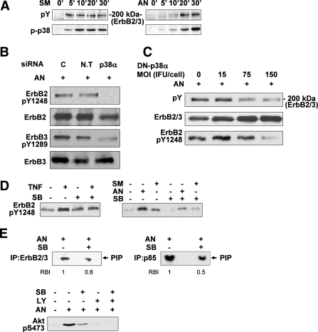
FIG. 4.
p38MAPK involvement in stress-induced ErbB2/ErbB3 transactivation in Fao cells. A: Cells were incubated with 300 mU/mL SMase or 50 ng/mL AN for the indicated times at 37°C. Total cell proteins were analyzed by SDS-PAGE, and the blots were probed with anti-pTyr or anti-p-p38 antibodies. B and C: Cells transfected by p38α or nontargeted (N.T) siRNA for 96 h (B) or infected with DN-p38α adenoviral constructs at the indicated MOI (ifu/cell) for 48 h (C) were treated with 50 ng/mL AN for 30 min. ErbB2-pTyr1248, ErbB3-pTyr1289, pTyr, ErbB2, and ErbB3 in total cell extract proteins were examined with immunoblot analysis using the indicated antibodies. The levels of ErbB2 and ErbB3 (ErbB2/3) in C, second panel, were assessed by immunoblot analysis using a mixture of anti-ErbB2 and anti-ErbB3 antibodies (2 μg each). D: Cells were treated without or with 10 μmol/L SB203580 (SB) for 30 min, before incubation without or with 5 nmol/L TNF, 300 mU/mL SMase, or 50 ng/mL AN for 30 min at 37°C. Total cell extract proteins were analyzed by Western immunoblot analysis using anti-ErbB2-pTyr1248 antibodies. E: Cells were treated with 10 μmol/L SB203580 (SB) and 50 ng/mL AN as described above. Immunoprecipitation was performed using p85-PI3K or mixture of anti-ErbB2 and anti-ErbB3 antibodies and PI3K activity was measured as described in research design and methods (E, upper panels). Cells were treated without or with 25 μmol/L LY294002 (LY) or 10 μmol/L SB203580 (SB) for 30 min before incubation without or with 50 ng/mL AN for 30 min. Akt activation was analyzed by immunoblotting with anti-Akt-pSer473 antibodies (E, lower panel). Each panel in A–E contains a representative blot of at least three independent experiments. The RBI of a given band in E, upper panels, presents the means of three independent experiments. Band intensity measurements were performed using ImageJ analysis.

