FIG. 5.
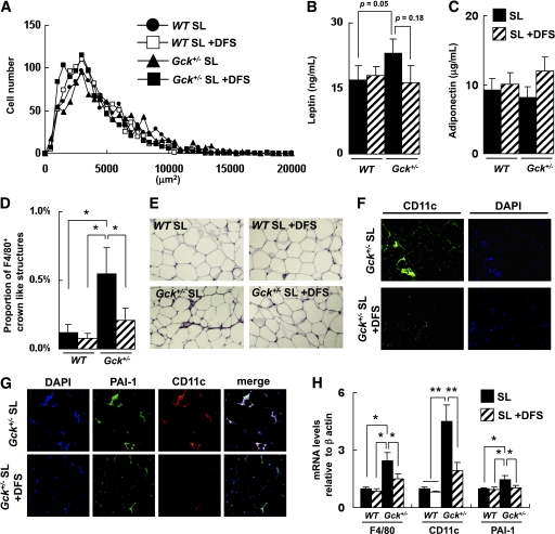
DFS prevented SL-induced adipose tissue infiltration by M1 macrophages and expression of PAI-1. A: Histogram of adipocyte size of the epididymal fat (n = 5). B and C: Serum leptin and adiponectin levels (n = 6–8). D and E: Epididymal fat tissue was stained with anti-F4/80 antibody. The number of CLSs was counted as described in research design and methods (n = 5). F: Epididymal fat tissue was stained with anti-CD11c antibody and DAPI. G: Epididymal fat tissue was stained with anti–PAI-1 antibody and anti-CD11c antibody. Experiments were performed on wild-type (WT) and Gck+/− mice after 25 weeks on the SO or SL diet. H: Assessment of the level of expression of the mRNAs indicated in epididymal fat as determined by real-time quantitative RT-PCR and normalization to the β-actin mRNA level (n = 5). Experiments were performed on wild-type and Gck+/− mice after 20 weeks on the SL or the SL-plus-DFS diet. *P < 0.05. **P < 0.01. (A high-quality digital representation of this figure is available in the online issue.)

