FIG. 7.
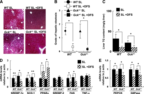
DFS protected against diet-induced hepatic steatosis. A: Masson-Goldner staining of liver sections from the groups of mice indicated. B: Grade of hepatic steatosis (grade 0–3) as described in Supplementary Fig. 14 (n = 7). C: Concentration of liver triglyceride in the groups of mice indicated (mg/g tissue) (n = 6). D: Hepatic gene expression of SREBP-1c, SCD-1, PPARα, SREBP-2, FAS, and TNF-α in the groups of mice indicated normalized to the β-actin mRNA level (n = 6). E: Hepatic gene expression of PEPCK and G6Pase in the groups of mice indicated normalized to the β-actin mRNA level (n = 5). Experiments were performed on wild-type (WT) and Gck+/− mice after 20 weeks on the SL diet or SL-plus-DFS diet. *P < 0.05. **P < 0.01. (A high-quality digital representation of this figure is available in the online issue.)

