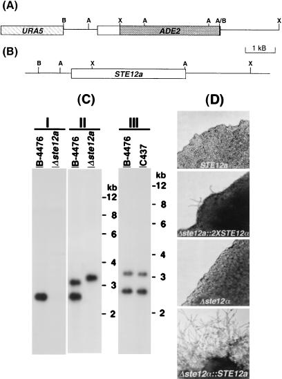
Figure 2.
Deletion and reconstitution of STE12a. (A) Map of the deletion plasmid construct, pYCC384. (B) Map of STE12a reconstitution construct, pYCC367. A, AseI; B, BamHI; X, XbaI. (C) Southern blot analysis of the Δste12a strain (I and II) and STE12a reconstituted strain (III). DNA was digested with AseI, fractionated on an agarose gel, and transferred to a nylon membrane. The resulting blot was hybridized with 2.1-kb XbaI–AseI probe of STE12a (I) or 3.6-kb XbaI–XbaI probe of STE12a (II and III). Wild type, B-4476; Δste12a, TYCC384; Δste12∷STE12a, C437. (D) Hyphae formation on filament agar. Strains B-4476, C490, TYCC245F1, and C489 were inoculated on filament agar. Photographs were taken after 3 days of incubation at room temperature.

