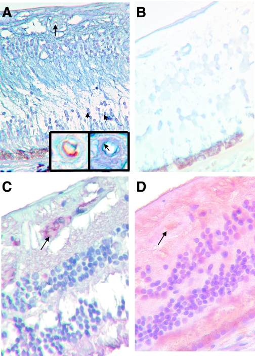
FIG. 6.
miR-200b alteration present in human diabetic retinopathy. A: LNA-ISH study of retinal tissues from nondiabetic human retina shows localization of miR-200b in the cells of the inner nuclear layer (arrowheads) and retinal capillaries (arrow). The inset (right) shows an enlarged view of the same capillary with endothelial localization of miR-200b (blue chromogen, arrow) and CD34 stain (brown chromogen) from an adjacent section showing endothelium of the capillary. B: Retinal tissues in a diabetic human retina (in similar orientation) show minimal (if any) expression of miR-200b. C: Immunocytochemical stain on the nondiabetic human retina using antialbumin antibody shows intravascular albumin (arrow). D: Diabetic human retina showed intravascular albumin staining (arrow) and diffuse staining of the retina, indicating increased vascular permeability. (ALK Phos was used as chromogen [blue] with no counterstain in LNA-ISH; DAB chromogen [brown] and hematoxylin counterstain in albumin stain.) (Original magnification ×400.) (A high-quality digital representation of this figure is available in the online issue.)

