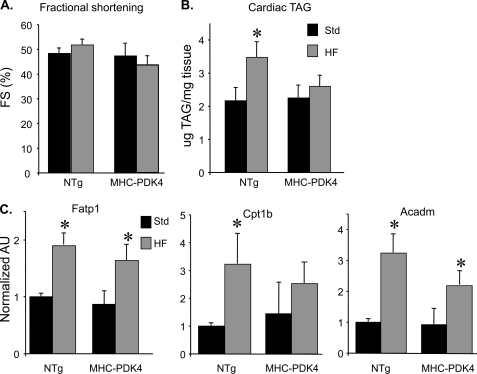
FIGURE 4.
MHC-PDK4 mice are protected from HF diet-induced TG accumulation in the myocardium. A, echocardiography was performed on male NTg and MHC-PDK4 mice (n = 4–6 per group after standard or high fat chow diet). Bars represent mean percent left ventricular fractional shortening (FS) % (±S.E.) after 4 weeks of HF diet (gray bars) versus standard chow (black bars). B, bars denote mean cardiac triglyceride (TG) levels (± S.E.) in male NTg and MHC-PDK4 hearts after 4 weeks of either standard (black bars) or HF chow (gray bars) (n = 6–9 per group). C, levels of mRNAs as determined by quantitative real-time-PCR were performed on total RNA isolated from heart ventricles of male mice from the genotypes indicated (n = 6–9 per group). Values are shown as arbitrary units (AU) normalized (=1.0) to the NTg on standard chow. *, p < 0.05.

