FIGURE 1.
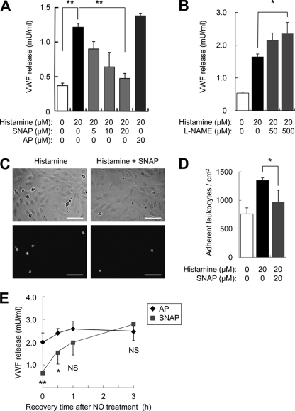
Nitric oxide inhibition of exocytosis is reversible. A, NO inhibition of VWF release. HUVEC were pretreated with the NO donor SNAP or its control AP for 6 h, stimulated with histamine, and the amount of VWF released into the media over 45 min was measured by an ELISA (n = 3, mean ± S.D.). The NO donor SNAP decreases endothelial exocytosis. B, endogenous NO inhibits VWF release. HUVEC were pretreated with the NOS inhibitor l-NAME for 4 h, stimulated with histamine, and the VWF released into the media was measured as above (n = 3, mean ± S.D.). The NOS inhibitor l-NAME increases endothelial exocytosis. C, NO inhibition of leukocyte adhesion. HUVEC were pretreated with SNAP for 4 h, stimulated with histamine for 20 min, then co-cultured with calcein-labeled HL-60 cells for 15 min. The upper panels show bright field images of HUVEC and HL-60, and the lower panels show calcein-labeled HL-60 co-cultured with HUVEC. Scale bars = 100 μm. D, quantitation of NO inhibition of leukocyte adhesion in C (n = 3, mean ± S.D.). The NO donor SNAP decreases leukocyte adhesion to endothelial cells. E, NO inhibition of exocytosis is reversible. HUVEC were pretreated with SNAP (20 μm) for 6 h, and then after recovery for 0, 0.5, 1, or 3 h the cells were stimulated with histamine (20 μm). Exocytosis was measured as above (n = 4, mean ± S.D.). The effect of NO upon exocytosis diminishes over time. *, p < 0.05; **, p < 0.01; NS, not significant.

