FIGURE 2.
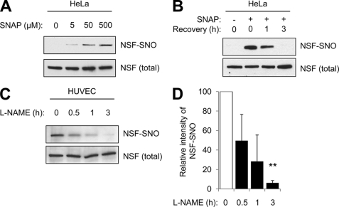
S-nitrosylation of NSF is reversible. A, S-nitrosylation of NSF in HeLa cells. HeLa cells were treated with the NO donor SNAP for 6 h. Then, cell lysates were harvested and levels of NSF-SNO were measured by the biotin switch assay. The NO donor SNAP increases NSF-SNO in a dose-dependent manner. B, S-nitrosylation of NSF decreases over time in HeLa cells. HeLa cells were treated with SNAP for 6 h and then washed. After 0–3 h of recovery, cell lysates were harvested, and levels of NSF-SNO were measured by the biotin switch assay. After exposure to an NO donor, the amount of S-nitrosylated NSF decreases over time. C, endogenous S-nitrosylation of NSF decreases over time in endothelial cells. HUVEC were treated with the NOS inhibitor l-NAME for 0–3 h, and then levels of NSF-SNO were measured as above. After NO synthase is inhibited, the amount of endogenous S-nitrosylated NSF decreases over time. D, quantitation of C (mean ± S.D). **, p < 0.01.

