FIGURE 2.
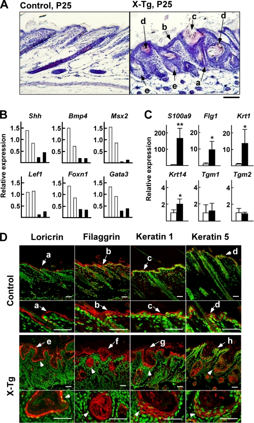
Hair follicle distortion, epidermal hyperplasia with hyperkeratosis, and sebaceous gland enlargement in the skin of PLA2G10-Tg mice. A, sections from control and PLA2G10-Tg (X-Tg) mice at P25 were stained with hematoxylin and eosin. In the Tg mice, the hair shafts appeared thin and distorted (arrow a); epidermal thickening (arrow b) and enlarged hair follicles (arrow c) were obvious; some follicles had degenerated into cyst-like structures filled with keratinized debris and surrounded by multilayered epithelium (arrow d); sebaceous glands displayed hyperplasia (arrow e). B and C, expression of genes associated with the differentiation of hair follicles and hair shafts (B) and of the interfollicular epidermis (C) in the skins of PLA2G10-Tg mice (solid columns) and control mice (open columns) at P25 was assessed by quantitative RT-PCR. Data are two representative results for each genotype (B) and means ± S.D. (n = 4; *, p < 0.05; **, p < 0.01) (C). The expression levels of individual genes in control mice are regarded as 1, with the expression of ribosomal RNA (18 S) as a reference. D, confocal laser scanning microscopy of various epidermal differentiation markers in dorsal skins from control and X-Tg mice at P25 is shown. Individual markers were stained red with specific antibodies, and nuclei were stained green with DAPI. Loricrin was present in the cornified layers (arrows a and e), filaggrin in the cornified and granular layers (arrows b and f), keratin 1 in the granular and spinous layers (arrows c and g), and keratin 5 in a single basal layer (arrows d and h). In X-Tg mice, loricrin-, filaggrin-, keratin 1-, and keratin 5-positive signals were distributed in the cysts (arrowheads). Bars represent 20 μm for all panels.

