FIGURE 2.
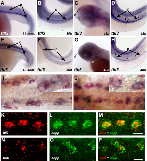
Expression of ttll3 and ttll6 is enriched in ciliated tissues. A–D, RNA in situ hybridization showing ttll3 is expressed during early development in distinct ciliated tissues: A, in the presumptive pronephros (p) at the 18-somite stage; B, at 30 hpf in discrete cells of the pronephros and at 48 hpf in olfactory placode (C) (n); D, lateral line organs (arrowheads) and in the pronephros (p; arrows). E–H, RNA in situ hybridization showing ttll6 is expressed in distinct ciliated tissues: E, the presumptive pronephros of an 18-somite embryo; F, in discrete cells of the pronephros at 30 and 48 hpf (G) in the olfactory placode; H, lateral line organs (arrowheads) and the pronephros (p). I and J, bright field images of two-color RNA in situ hybridization, showing transcripts of both ttll3 (I) (blue) and ttll6 (J) (blue) , respectively, colocalized with shpp (red), a marker of multiciliated cells. Insets within each panel show individual cells at higher magnification. K–P, fluorescent two-color RNA in situ hybridization confocal images showing antisense probes of ttll3 (red) (K); ttll6 (red) (N) and the multiciliated cell marker shpp (green) (L and O) in a single confocal slice of the pronephros (30 hpf). In the merged panels, coexpression of ttll3 (M) with shpp (green) (red) and ttll6 (red) with shpp (green) (P) indicates that transcripts of both ttll3 and ttll6 are enriched in multiciliated cells. Scale bars in M and P = 10 μm.

