FIGURE 3.
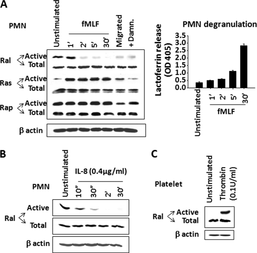
Ral deactivation correlates secondary granule release in PMN. A, detecting activity changes of Ras family small GTPases, including Ral, Ras, and Rap, in PMN. After time course stimulation with fMLF (1 μm), PMN were lysed, and active Ras, Ral, and Rap were pulled down using Raf-1 RBD, RalBP1, and RalGDS-RBD, respectively. Right, in parallel, fMLF-induced PMN degranulation of secondary granules was assayed simultaneously by detecting lactoferrin in the cell-free supernatants. PMN that transmigrated across collagen-coated filters into the fMLF-containing lower chambers or PMN that were treated with damnacanthal were also assayed for active Ral, Ras, and Rap. Cell lysates before pull-down were used to detect total Ral, Ras, and Rap as well as actin using antibodies against Ral, Ras, Rap, and β-actin. B, deactivation of Ral by IL-8 stimulation. Recombinant IL-8 (0.4 μg/ml) (Sigma) was used to stimulate PMN for different time periods, followed by RalBP1 pull-down to detect active Ral. C, Ral activities in unstimulated and thrombin (0.1 unit/ml) (Sigma)-stimulated platelets. Data (mean ± S.D. (error bars)) represent three independent experiments with triplicates in each condition.

