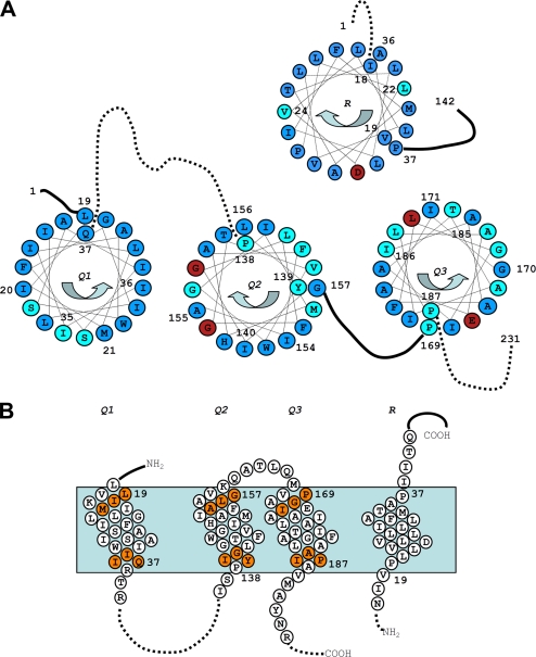
FIGURE 2.
Summary of the TolQ cysteine substitution phenotypes. A, TolQ TMHs residues shown on a helical wheel projection from the periplasmic to the cytoplasmic side. The color code summarizes the result of the phenotypic analyses of the cysteine substitutions (Table 1): WT phenotype (blue, colicin-sensitive and DOC-resistant), tol phenotype (red, colicin-resistant and DOC-sensitive), and discriminative phenotype (cyan, colicin- and DOC-sensitive). The residues used in the tandem cysteine scanning and Ile154 are numbered. Residues numbered on the outside of the wheel locate at the periplasmic side of the TMH, whereas residues numbered on the inside locate at the cytoplasmic side. Periplasmic and cytoplasmic segments are indicated by unbroken and dashed lines, respectively. The phenotypic analysis of TolR TMH cysteine substitutions (30) is also reported using the same color code. B, schematic lateral view of TolQ and TolR TMHs in which the residues used for the tandem cysteine mutagenesis are indicated by orange balls. The residues locating at the boundaries of each TMH are numbered.

