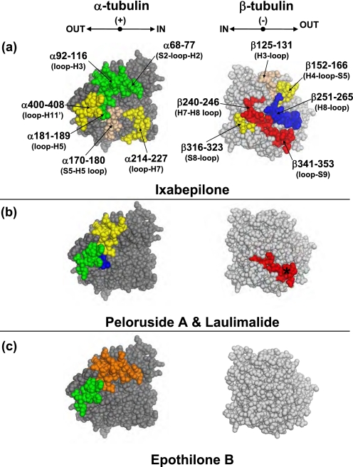
FIGURE 5.
Mapping the local HDX alteration on the intradimer interface of the tubulin dimer (PDB code 1JFF). Peptides are colored according to the code in Fig. 4. In addition, blue = significant increase in deuterium incorporation (ΔHDX > 0). The conformational effects of ixabepilone (a), peloruside A and laulimalide (b), and epothilone B (c) are illustrated. In b the asterisk marks peptide β341–353, which is strongly protected by all drugs but significantly more so by peloruside A and laulimalide. For clarity, only regions significantly different from panel a are shown in color in panels b and c. Secondary structure designations are based on Löwe et al. (6) The directional coordinates are shown in a above each α- and β-tubulin component of the interface, with designations as indicated in Figs. 3 and 4.

