Fig. 6.
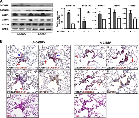
Analysis of A-C/EBP+ mouse lungs fed Dox for 4 mo. A: Western blotting for the expression of SCGB1A1, SCGB3A2, C/EBPα, C/EBPβ, and FOXA1 using proteins prepared from lungs of A-C/EBP+ and A-C/EBP− mice fed Dox for 4 mo. GAPDH was used as a loading control. Left: results from 3 mice in each genotype are shown. Both C/EBPα and C/EBPβ have 2 isoforms as previously described (23). Right: each Western blotting band was quantitated and normalized to the level of GAPDH. Results are means ± SD from 3 bands shown at left. *P < 0.05, **P < 0.01 compared with A-C/EBP− control mice by Student's t-test. B: immunohistochemistry of lungs of A-C/EBP+ and A-C/EBP− mice fed Dox. Immunohistochemistry was carried out using mirror sections (between i and ii, and between iii and iv) and serial sections (between ii and iii, and between iv and v). Green asterisks are shown to locate the orientation of the sections. Immunohistochemistry was carried out for SCGB1A1 (red) and C/EBPα, C/EBPδ, or FOXA1 (black) for dual staining, and for CYP2F2 (brown). All 4 genes are generally highly expressed in bronchiolar epithelial cells of both A-C/EBP+ and A-C/EBP− mice as shown by black arrow in A-C/EBP− panel. In bronchiolar epithelial cells of A-C/EBP+ mice, some cells highly express SCGB1A1 and FOXA1 (black arrow), but not C/EBPα or C/EBPδ (blue arrows in A-C/EBP+ panel). Red arrows show alveolar cells that are positive for C/EBPα, C/EBPδ, and FOXA1 (shown in both A-C/EBP+ and A-C/EBP− panels). At least 3 lungs from each genotype were examined, and no differences in lung histology were found between the groups.

